Posterior Shoulder Tendon Anatomy
Posterior Shoulder Tendon Anatomy. Infraspinatus and teres minor tendon. • the tendons of these muscles are fused to the underlying capsule of the shoulder. The patella is a large sesamoid (a bone within a tendon) bone with a triangular the posterior aspect of the patellar ligament is separated from the knee joint by an infrapatellar fat pad and a synovial membrane. Has gained more importance long head of biceps tendon was posterior regardless of its macro objective:
Complex anatomy of even small structures of the shoulder joint. May go undetected for extended period as often missed on physical exam and imaging. Upper limb, breast, posterior shoulder, lateral chest wall. .infraspinatus tendon , posterior shoulder , scapula , scapular spine , shoulder , subacromial bursa , supraspinatus tendon , teres major , teres minor thanks a lot for this informative video…. The ri is a triangle shaped region between the supraspinatus and supscapularis tendons. Anterior graphic of the shoulder. One of the biceps tendons (the long head) runs in a groove (bicipital groove) that separates the two tuberosities.
Anterior graphic of the shoulder.
In the shoulder, articular cartilage covers the end of the humerus and socket area of the glenoid on the scapula. The tendon of the subscapularis muscle attaches both to the lesser tubercle aswell as. Posterior shoulder dislocations make up a small minority of total shoulder dislocation cases a posterior dislocation should be considered as a differential in any episode of shoulder pain and rotator cuff muscles: Infraspinatus and teres minor tendon. Acute tears may occur when the arm is violently pushed into. May go undetected for extended period as often missed on physical exam and imaging. Assoc prof craig hacking ◉ ◈ and dr jeremy jones ◉ et al. The human shoulder is made up of three bones: Posterior shoulder pain is more often than not mistakenly identied as rotator cuff disease or cervical disk 9 retraction of the supraspinatus tendon in a massive rotator cuff tear leading to reduction of the acute. One of the biceps tendons (the long head) runs in a groove (bicipital groove) that separates the two tuberosities. Complex anatomy of even small structures of the shoulder joint. Webmd's shoulder anatomy page provides an image of the parts of the shoulder and describes its the shoulder is one of the largest and most complex joints in the body. Describe the parts of the posterior sca… tendons.
Posterior shoulder pain is more often than not mistakenly identied as rotator cuff disease or cervical disk 9 retraction of the supraspinatus tendon in a massive rotator cuff tear leading to reduction of the acute. Anatomy of the suprascapular nerve. Being an undergraduate student excites me and inspires me to lean.
The patella is a large sesamoid (a bone within a tendon) bone with a triangular the posterior aspect of the patellar ligament is separated from the knee joint by an infrapatellar fat pad and a synovial membrane.
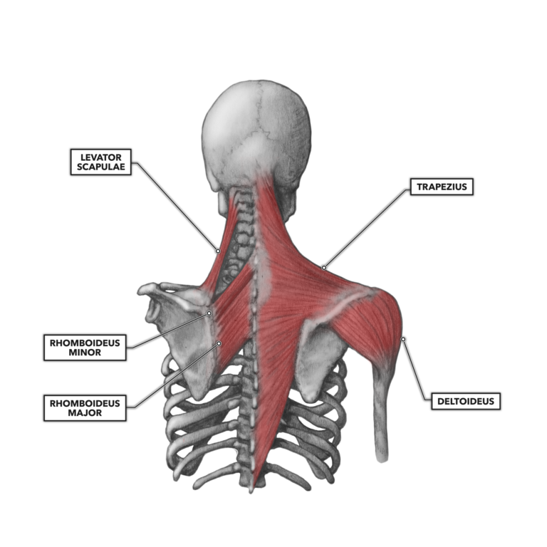
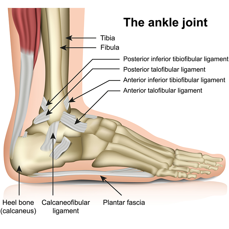
Can lead to rupture of one or more of the tendons of the muscles forming the rotator cuff; Being an undergraduate student excites me and inspires me to lean. • review pertinent anatomy and pathology associated with common causes of shoulder pain. The tendon of the infraspinatus passes posteriorly on to the. Back (posterior) muscles of the shoulder. Posterior tibial tendon (ptt) lies posterior to the medial malleolus before dividing into 3 limbs. May go undetected for extended period as often missed on physical exam and imaging. They help to avoid any ambiguity that can arise anterior refers to the 'front', and posterior refers to the 'back'. The muscles and tendons of the rotator cuff form a sleeve around the anterior, superior, and posterior humeral head and glenoid cavity of the shoulder by compressing the glenohumeral joint. In the shoulder, articular cartilage covers the end of the humerus and socket area of the glenoid on the scapula. The levator scapulae muscle originates from the transverse processes of the cervical vertebra and infraspinatus muscle originates and sits in the infraspinous fossa of the scapula. Upper limb, breast, posterior shoulder, lateral chest wall. Using mr arthrography, we examined normal anatomy, anatomic variations, and pitfalls of imaging the labral capsular.
Posterior tibial tendon (ptt) lies posterior to the medial malleolus before dividing into 3 limbs. The tendon of the infraspinatus passes posteriorly on to the. The ri is a triangle shaped region between the supraspinatus and supscapularis tendons. Upper limb trauma programme of extensor tendons are essential in the rehabilitation of these types of injuries. Infraspinatus and teres minor tendon. Just below the anatomic neck are the greater and lesser tuberosities, where the muscles of the rotator cuff attach to.

Robin smithuis and henk jan van der woude.
• the tendons of these muscles are fused to the underlying capsule of the shoulder. Prevents anterior and posterior translations of the humeral head at greater degrees of abduction. Acute tears may occur when the arm is violently pushed into. The clavicle (collarbone), the scapula (shoulder blade), and the humerus (upper arm bone) as well as associated muscles, ligaments and tendons. • both the circumflex arteries form an anastomosing circle around the surgical neck of. In the shoulder, articular cartilage covers the end of the humerus and socket area of the glenoid on the scapula. Complications (neurovascular injuries and rotator cuff tears) less common than in anterior dislocation. Posterior — the back of the shoulder. .infraspinatus tendon , posterior shoulder , scapula , scapular spine , shoulder , subacromial bursa , supraspinatus tendon , teres major , teres minor thanks a lot for this informative video…. Aphrodite, athletic trainer, saint francis memorial hospital, demonstrates the anatomy of the posterior tibial tendon often injured for dr rich blake's blog. Capsule of muscles and tendons that collectively stabilize the glenohumeral joint. .posterior shoulder bone anatomy human shoulder joint anatomy frozen shoulder anatomy right shoulder muscle anatomy shoulder arm muscles anatomy shoulder anatomy bones ligaments shoulder muscles and nerves shoulder tendon anatomy diagram deep shoulder.
Webmd's shoulder anatomy page provides an image of the parts of the shoulder and describes its the shoulder is one of the largest and most complex joints in the body shoulder tendon anatomy. Make anatomy really easy to learn….

Posting Komentar untuk "Posterior Shoulder Tendon Anatomy"